New Statistical Models Improve Multiplexed Imaging Analysis
A new publication in Cell Systems from the Bodenmiller lab describes innovative statistical models designed to improve the analysis of cell counts in multiplexed imaging data, an essential tool for precision medicine research.
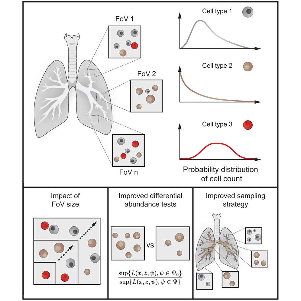
Multiplexed imaging allows detailed visualization of complex tissues, yet existing statistical methods to analyze cellular distribution and abundance often lack sensitivity. To address this, the team developed two probabilistic models—the negative binomial and beta binomial distributions—to accurately describe variations in cell counts across tissue samples. These models account for factors like field-of-view size, cellular density, and spatial aggregation patterns, significantly enhancing the accuracy and reliability of comparative tissue analysis.
The new methods substantially outperformed traditional rank-based statistical tests in identifying subtle but biologically meaningful differences in cell abundance. By improving statistical sensitivity, these models reduce the sample sizes needed, enhancing both efficiency and reliability for clinical studies of diseases like cancer and autoimmune conditions.
This advancement offers a powerful analytical toolset for researchers using multiplexed imaging, facilitating more accurate clinical insights and personalized therapeutic strategies.
Publication Link: https://doi.org/10.1016/j.cels.2025.101296