The Bodenmiller group develops a 3D IMC approach for multiplexed 3D tissue analysis at single-cell resolution
Three-dimensional imaging mass cytometry for highly multiplexed molecular and cellular mapping of tissues and the tumor microenvironment
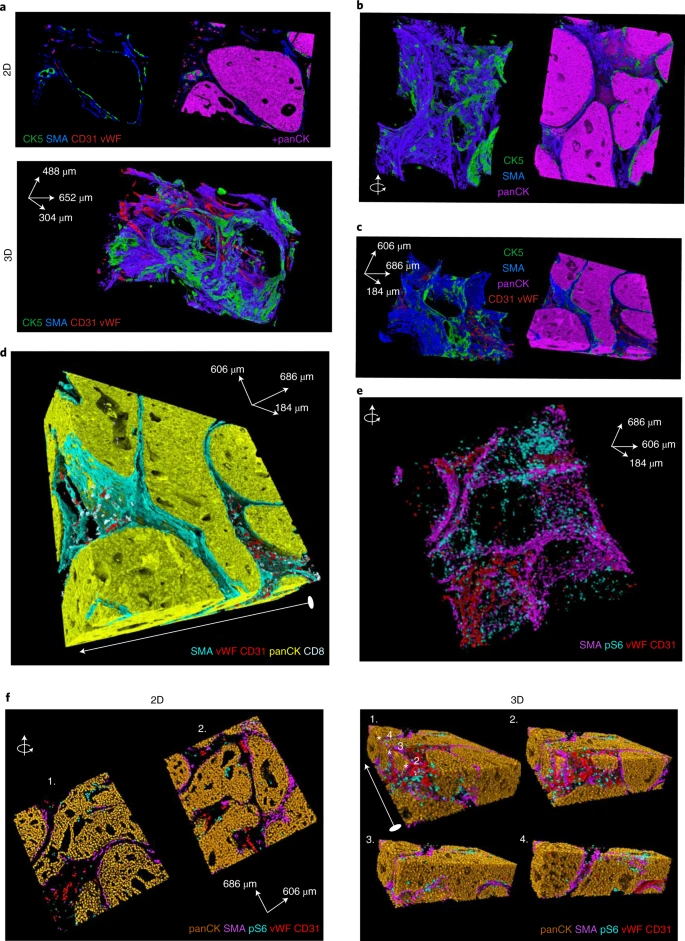
Understanding tissue function and pathology requires knowledge of the molecular components in their original three-dimensional (3D) context. In this work, the Bodenmiller group reports on the development of 3D Imaging Mass Cytometry (IMC) for multiplexed 3D tissue analysis and demonstrates, using the analysis of human breast cancer samples, that the detailed models generated by 3D IMC enable comprehensive analysis of the cellular microenvironment and tissue architecture at single-cell resolution. Thus, 3D-IMC advances the study of the spatial distribution of proteins in the context of 3D tissue architecture and will prove powerful in the study of phenomena such as tumor cell invasion.
See Kuett et al., Nature Cancer