Navigation auf uzh.ch
Navigation auf uzh.ch
To predict the effects of immunotherapy in cancer, better spatial understanding of the tumor microenvironment (TME) is needed.
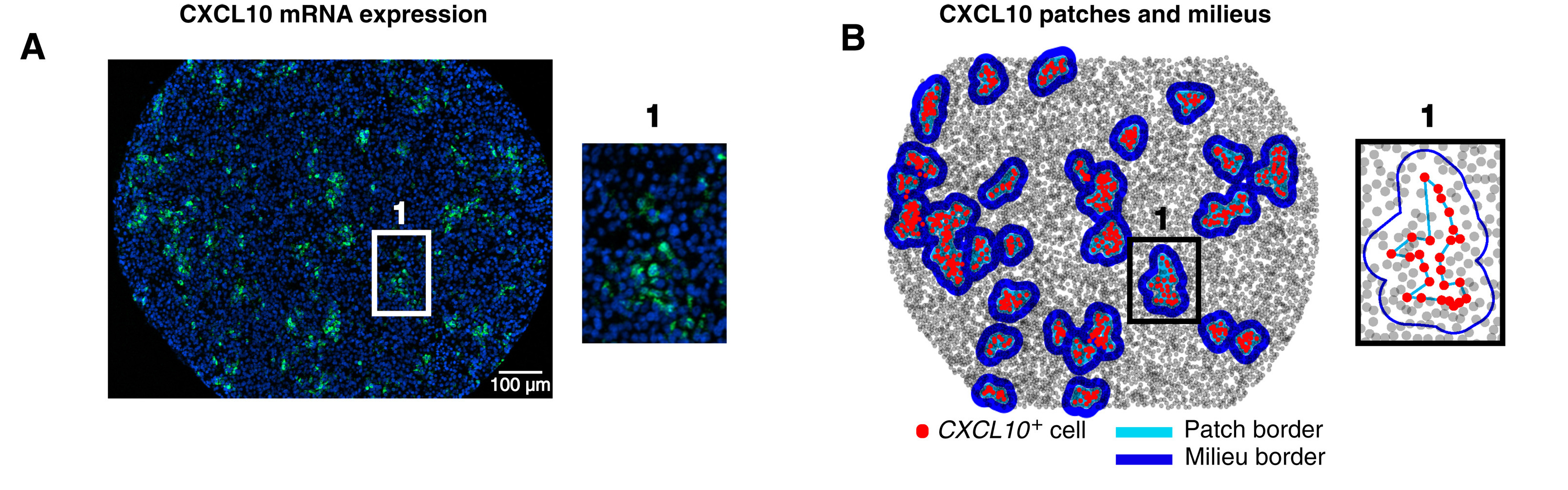
In this work, the Bodenmiller group characterised chemokine expression and function in samples from 69 patients with metastatic melanoma. Using multiplexed mass cytometry-based imaging of protein markers and RNA transcripts, they found that CXCL9 and CXCL10 were present in patches with CXCL13+ exhausted T cells, suggesting that they recruited B cells and aided in the formation of tertiary lymphoid structures (TLS) in melanoma. TLS had a spatial enrichment of naïve and naïve-like
T cells, which are involved in anti-tumor responses. Together, this study highlights the strength of targeted RNA and protein co-detection to analyse TME based on chemokine expression and suggests that the formation of tertiary lymphoid structures may be accompanied by naïve and naïve-like T cell recruitment, which may contribute to anti-tumor activity.
See Hoch, Schulz et al., Sci Immunol